

The effort follows Trump’s controversial proposal to “own” the Gaza Strip as part of a plan he has to establish “stability” in the Middle East—a move that would displace the two million Palestinians who reside there.
During a White House press conference on Tuesday (February 4), alongside Israeli Prime Minister Benjamin Netanyahu, the 47th U.S. President outlined his vision to “take over” the Gaza Strip, leaving the possibility of U.S. military involvement open.
“The only reason the Palestinians want to go back to Gaza is they have no alternative, it’s right now a demolition site,” Trump stated. “This is just a demolition site. Virtually every building is down.”
He also described his intention to relocate Palestinian residents to neighboring countries such as Egypt and Jordan.
“The U.S. will take over the Gaza Strip and we will do a job with it, too,” he continued. “We’ll own it and be responsible for dismantling all of the dangerous unexploded bombs and other weapons on the site.”
Attempting to cast his plan in a positive light, Trump argued that his administration would redevelop Gaza, claiming it would “create thousands and thousands of jobs, and it’ll be something that the entire Middle East can be very proud of.”
Trump’s announcement has sparked significant backlash, with human rights advocates condemning it as “ethnic cleansing by another name.”
Democratic Senator Chris Van Hollen of Maryland criticized the proposal, stating: “Trump’s proposal to push 2 million Palestinians out of Gaza and take ‘ownership’ by force, if necessary, is simply ethnic cleansing by another name.
“This declaration will give ammunition to Iran and other adversaries while undermining our Arab partners in the region. It defies decades of bipartisan American support for a two-state solution… Congress must stand up to this dangerous and reckless scheme.”
Texas Representative Al Green also denounced Trump’s plan, referring to Gaza as the “Riviera of the Middle East.”
“Ethnic cleansing in Gaza is not a joke, especially when it emanates from the President of the United States, the most powerful person in the world,” Green said. “And the prime minister of Israel should be ashamed, knowing the history of his people, to stand there and allow such things to be said.”
He invoked the words of Dr. Martin Luther King Jr., adding: “Dr. [Martin Luther] King was right. Injustice anywhere is a threat to justice everywhere, and injustice in Gaza is a threat to justice in the United States of America.”
Green then formally announced the start of impeachment proceedings: “I rise to announce that the movement to impeach the president has begun. I rise to announce that I will bring articles of impeachment against the president for dastardly deeds proposed, and dastardly deeds done.”
He further declared, “[The] impeachment movement is going to be a grass-up movement, not a top-down. When the people demand it, it will be done.”
Once Green submits the articles of impeachment, the House of Representatives will vote. If a majority supports impeachment, Trump will be formally impeached.
The case would then move to the Senate for a trial overseen by the U.S. Supreme Court’s chief justice.
Should Trump be found guilty, he would be removed from office.
However, political analysts suggest Green’s impeachment effort faces significant hurdles. House Democratic leadership does not appear to prioritize impeachment at this time, with Pete Aguilar, the No. 3 House Democrat, telling Politico that it is “not an immediate focus of his caucus.”
This is not Green’s first attempt to impeach Trump—he previously pushed for impeachment during Trump’s first term.
Trump has already made history as the first U.S. president to be impeached twice, in 2019 and 2021.


![Figure 1. Pincer nail of the left first toenail in an 80-year-old woman. The lateral aspect of the nail plate is penetrating the periungual dermis of the lateral nail fold [Citation8].](https://www.tandfonline.com/cms/asset/b371fbc1-b699-4db4-925f-9fd76a8005b3/iann_a_2336989_f0001_c.jpg)
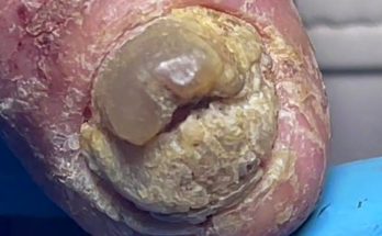
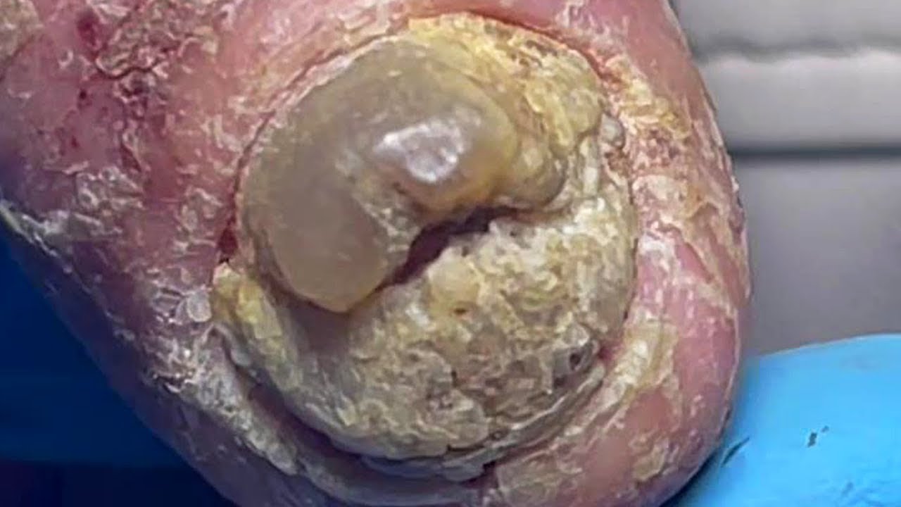
![Figure 2. A 75-year-old female presented with painful bilateral great toenails for 10 years. Her nails grew slowly and were extremely difficult to clip. A full nail examination was significant for opaque yellow-brown thickening, hyperkeratosis, elongation, and increased curvature of the great toenails. Onychogryphosis can be differentiated from retronychia and onychomycosis by its spiral striated appearance [Citation13].](https://www.tandfonline.com/cms/asset/37bfa230-31a9-4ea5-be2d-6213eb190967/iann_a_2336989_f0002_c.jpg)

![Figure 4. Clinical presentations of Beau’s lines, onychomadesis and retronychia. (A) Beau’s lines on the left toenails. (B) Onychomadesis of the left great toenail. (C) Retronychia of the right great toenail [Citation20].](https://www.tandfonline.com/cms/asset/28b7b61e-05eb-495a-8840-7852502e62e9/iann_a_2336989_f0004_c.jpg)
![Figure 5. 93-year-old female with bullous pemphigoid presented with Beau’s lines on all fingernails at even intervals coinciding with her monthly IVIG treatments [Citation23].](https://www.tandfonline.com/cms/asset/3aae46a7-e806-4a4b-8a85-7bb43bbf412a/iann_a_2336989_f0005_c.jpg)
![Figure 6. Physical examination findings in onychomycosis. A, Right great toenail with subungual hyperkeratosis and nail plate onycholysis. B, Left great toenail with yellow discoloration and onycholysis. C, Multiple toenails with subungual hyperkeratosis and onycholysis. D, Toenails with severe onychodystrophy and ridging. E, Scale on the plantar feet and web spaces [Citation33].](https://www.tandfonline.com/cms/asset/e92b5d45-1340-499a-b4bf-4f033e00cf45/iann_a_2336989_f0006_c.jpg)
![Figure 7. Dermoscopy of onychomycosis. A, Fringed proximal margin of the onycholysis. B, Blurred yellow-orange-brown nail discoloration in longitudinal striae (the fading mimics Aurora Borealis). C, Distribution of the discoloration in longitudinal striae or round areas. D, Ruin-like appearance of the subungual scales that are white-yellow-orange in color. Photographs courtesy of Dr Maria Bianca Piraccini [Citation34].](https://www.tandfonline.com/cms/asset/ebb4913a-53d9-49a3-aff4-ac5844a01c31/iann_a_2336989_f0007_c.jpg)


![Figure 10. Nail pitting and onycholysis in right fingernails [Citation20].](https://www.tandfonline.com/cms/asset/9660b1fb-2bbc-430f-9e27-9dcf72011b41/iann_a_2336989_f0010_c.jpg)
![Figure 11. Dermoscopic appearance of subungual melanomas. B, Brown lines on a brown background, irregular color, thickness, and spacing with no loss of parallelism. C, Brown lines on a brown background, irregular color, thickness, and spacing with loss of parallelism[Citation62].](https://www.tandfonline.com/cms/asset/74a5fabc-6954-4a49-83ce-75d6752880ac/iann_a_2336989_f0011_c.jpg)
![Figure 12. A, A translucent compressible nodule of the proximal nail fold and longitudinal groove in the nail plate of the right thumb. B, Transillumination using a dermatoscope to project light from the dorsal digit through the nail unit demonstrated a central nodule in the proximal nail fold as well as a second cyst radially. (Reprinted with permission from Cutis. 2020;105(2):82. ©2020, Frontline Medical Communications Inc.) [Citation73].](https://www.tandfonline.com/cms/asset/2e22d4d0-100e-4011-9cf8-89fbb8ab2381/iann_a_2336989_f0012_c.jpg)